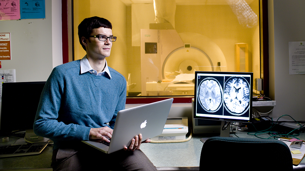
Andrew Walsh
As if becoming a physician wasn't hard enough.
As an engineering wunderkind, Andrew Walsh decided to apply for the MD/PhD program in 2008 after receiving his undergraduate degree.
Getting into the program is not easy: successful applications must already be accepted into the medical program-no easy undertaking-and the student then needs to be prepared to complete the first two years of medical school before entering the PhD program, before returning back into the medical milieu.
Only a handful of students have been accepted into the program each year since its inception in 2002, and only 26 students have successfully completed the program. The program is highly competitive, with only two or three students entering the program each year.
A daunting prospect for many, but not for Walsh, who graduates on June 5 and will be entering the radiology residency program in the Faculty of Medicine & Dentistry.
"The opportunity to marry my engineering interests within medicine seemed like a perfect fit to me," says Walsh about completing his undergraduate, post-graduate and medical training all at the University of Alberta. "The opportunities were all in the right place, at the right time. Biomedical engineering and medicine compliment my interest in engineering," he says.
"The MD/PhD program is a shining example of the competitive yet collegial environment we cultivate in the faculty," says Faculty of Medicine & Dentistry interim dean Richard Fedorak. "Andrew, like the MD/PhD graduates before him and those to follow, exemplify the very best in graduate research and medical education."
Walsh, who was selected for one of Canada's most prestigious graduate scholarships in 2010 when he was appointed as a Vanier Scholar, has focused that passion on advancing magnetic resonance imaging (MRI), a sophisticated imaging technique used in disease diagnosis, including multiple sclerosis (MS). Instead of using this tool to just diagnose MS patients by analyzing brain lesions-an important diagnostic methodology-Walsh conceived of a new idea: using MRI to measure iron levels in the brains of MS patients over a period of time to track disease progression.
The reason why iron levels in the brain of patients with MS are higher is not known, but Walsh's study shows a "distinct correlation" between brain iron content and the progression of the disease. Having made that discovery, Walsh then hypothesized that, by developing a standard of longitudinal assessment of these levels, the MRI could possibly be used as a compliment to drug treatments, providing a clear assessment to physicians about their patients' status and whether their treatments are actually effective.
"This is the first study of its kind, looking at the deep gray matter of the brain and its iron storage distribution as a marker of brain disease," says Walsh's PhD supervisor Alan Wilman, associate chair of the Department of Biomedical Engineering. "Andrew's plan, to look at iron levels over time to measure the amount of damage to the brain, is reflective of his determination and motivation.
"He pushed this forward very quickly and made great breakthroughs."
Walsh plans on building upon the four articles that he and Wilman authored during his PhD.
"My end goal is to work hand in glove with other health professionals to diagnose and treat neurological disorders," he says. With the combination of medical and research skills Walsh has gained as part of the elite MD/PhD program, Wilman is confident he has trained an emerging leader in his field.
"Andrew now has the scientific knowledge that will allow him to make breakthroughs, combined with a depth of medical knowledge that will enable him to understand the health implications of his own research."